
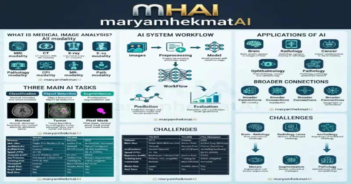
How AI and Computer Vision are used for classification, object detection, and segmentation of medical images
Medical images contain valuable information that can support healthcare research, diagnosis, and monitoring. However, analyzing large numbers of images manually can be time-consuming
Artificial Intelligence (AI), especially Machine Learning and Deep Learning, provides tools for analyzing medical images and identifying visual patterns.
Today, Computer Vision is being explored for tasks such as image classification, object detection, and image segmentation.
What Is Medical Image Analysis?
Medical image analysis is the use of computational methods to extract useful information from images such as:
MRI
CT scans
X-rays
Ultrasound
Mammography
Retinal images
Digital pathology images
Depending on the task, an AI model may classify an image, locate an abnormality, or identify specific regions at the pixel level.
For general biomedical research resources, see the National Institutes of Health (NIH)
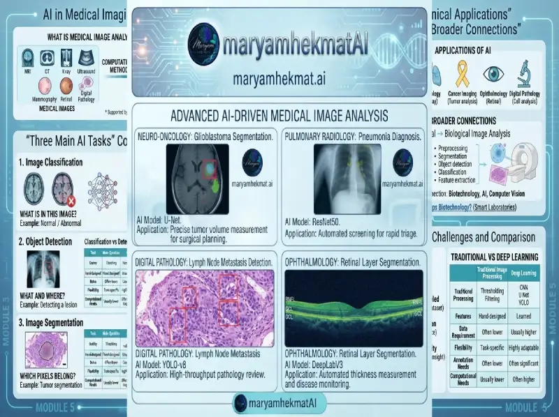
Three Main AI Tasks in Medical Imaging
Image Classification
Classification answers:
What is in this image?
For example, a model may classify an image as normal or abnormal.
Deep learning models, including CNN-based architectures, can learn visual patterns from labeled datasets.
Object Detection
Object detection answers two questions:
What is it?
Where is it?
Object detection methods such as YOLO-based models can be used for fast computer vision applications, including real-time object detection.
Image Segmentation
Segmentation works at the pixel level.
For example, a model can identify the exact region of a tumor, organ, cell, or other structure.
One well-known biomedical segmentation architecture is U-Net.
These three tasks are closely related, but they provide different levels of information.
How Does an AI Medical Imaging System Work?
A simplified workflow looks like this:
Medical Images → Preprocessing → AI Model → Prediction → Evaluation
Preprocessing may include:
- Resizing
- Normalization
- Noise reduction
- Contrast adjustment
- Data augmentation
The model then learns patterns from training data and produces predictions on new images.
Applications of AI in Medical Imaging
🧠 Brain Imaging
AI can be used to analyze MRI and CT images and study brain structures or detect specific patterns.
🫁 Radiology
Computer vision methods can be applied to X-rays and other radiological images for research and automated image analysis.
🎗️ Cancer Imaging
AI is being studied for tumor detection, classification, and segmentation.
👁️ Ophthalmology
Retinal images can be analyzed using machine learning and deep learning techniques.
🔬 Digital Pathology
Microscopic tissue images can be analyzed for cell detection, tissue classification, and quantitative measurements.
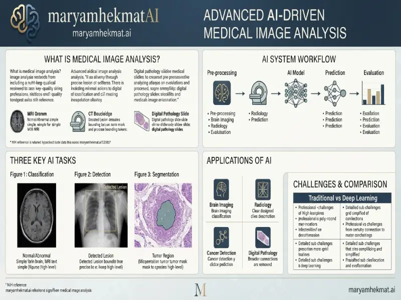
Medical Imaging and Biological Image Analysis
Medical image analysis has a strong connection with Biological Image Analysis and Microscopy Image Analysis.
Medical imaging may involve MRI, CT, or X-ray images, while biological imaging can involve cells, tissues, microorganisms, and microscopic structures.
Many of the same Computer Vision techniques can be used in both areas:
- Image preprocessing
- Segmentation
- Object detection
- Classification
- Feature extraction
This connection between Biotechnology, AI, and Computer Vision is one of the areas I am particularly interested in exploring.
You can also read my article How Does Artificial Intelligence Help Biotechnology? From Laboratory Image Analysis to Smart Laboratories.
Challenges of AI in Medical Image Analysis
AI can provide powerful tools, but medical imaging also presents important challenges.
Data Quality
Images can vary in resolution, quality, and acquisition methods.
Limited Labeled Data
Creating accurate medical annotations can require significant time and domain expertise.
Generalization
A model trained on one dataset may not perform identically on images from another hospital, device, or population.
Interpretability
Understanding why an AI model produced a particular prediction can be important in healthcare applications.
Privacy
Medical images and patient information require appropriate privacy and data protection practices.
Traditional Image Processing vs Deep Learning
Both traditional image processing and deep learning can be useful.
Leave a Reply