Author: admin

  • Medical Image Analysis Using Artificial Intelligence

    Medical Image Analysis Using Artificial Intelligence

    How AI and Computer Vision are used for classification, object detection, and segmentation of medical images

    Medical images contain valuable information that can support healthcare research, diagnosis, and monitoring. However,    analyzing large numbers of images manually can be time-consuming

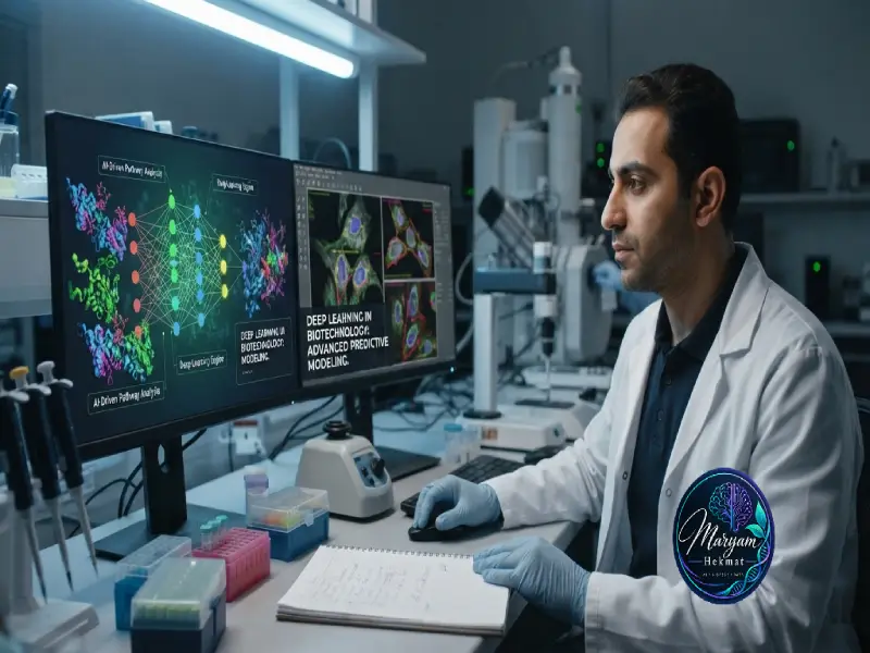
    Artificial Intelligence (AI), especially Machine Learning and Deep Learning, provides tools for analyzing medical images and identifying visual patterns.

    Today, Computer Vision is being explored for tasks such as image classification, object detection, and image segmentation.

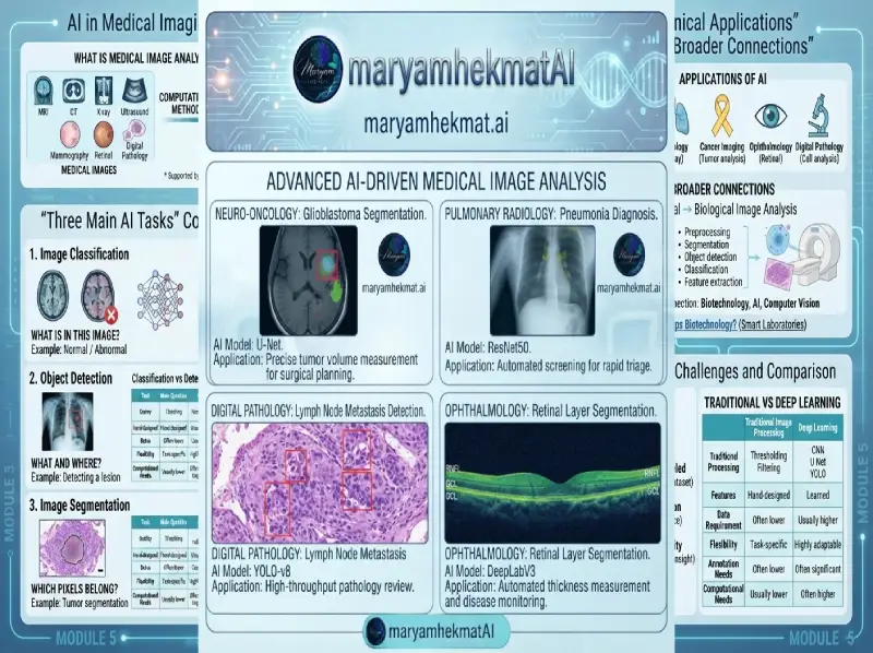
    What Is Medical Image Analysis?

    Medical image analysis is the use of computational methods to extract useful information from images such as:

    MRI

    CT scans

    X-rays

    Ultrasound

    Mammography

    Retinal images

    Digital pathology images

    Depending on the task, an AI model may classify an image, locate an abnormality, or identify specific regions at the pixel level.

    For general biomedical research resources, see the National Institutes of Health (NIH)
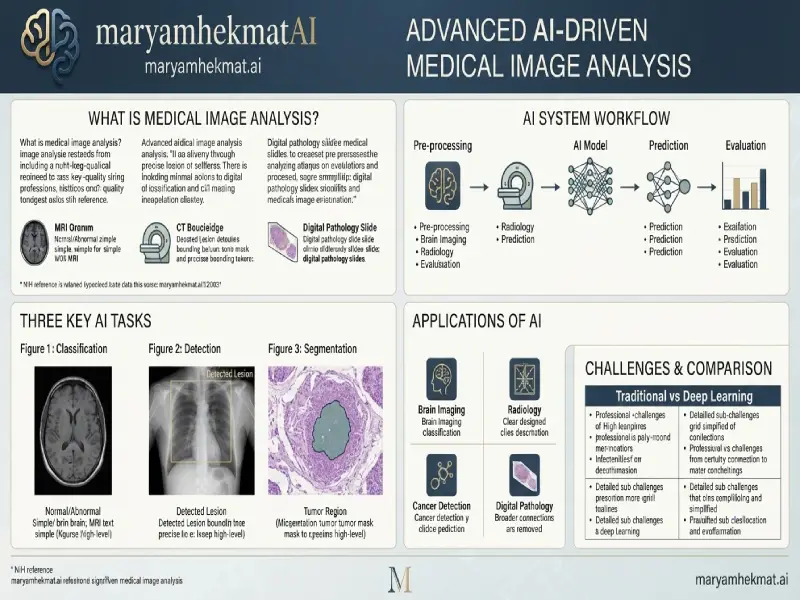
    How Does an AI Medical Imaging System Work_

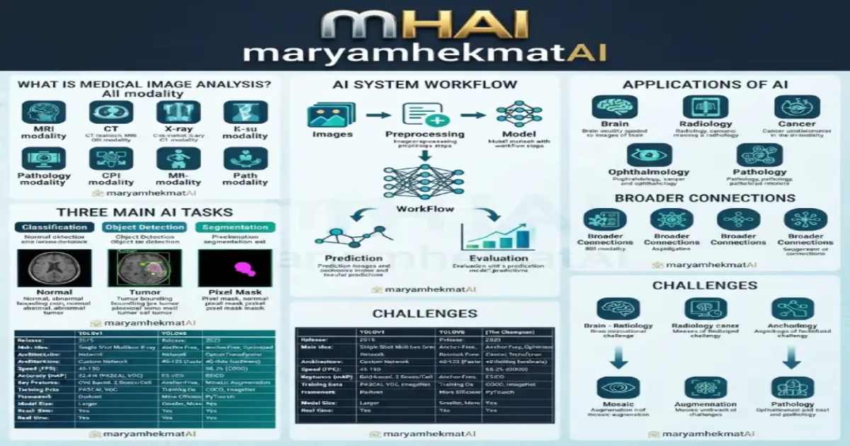
    Three Main AI Tasks in Medical Imaging


    1. Image Classification

    Classification answers:

    What is in this image?

    For example, a model may classify an image as normal or abnormal.

    Deep learning models, including CNN-based architectures, can learn visual patterns from labeled datasets.

    1. Object Detection

    Object detection answers two questions:

    What is it?
    Where is it?

    Object detection methods such as YOLO-based models can be used for fast computer vision applications, including real-time object detection.

    1. Image Segmentation

    Segmentation works at the pixel level.

    For example, a model can identify the exact region of a tumor, organ, cell, or other structure.

    One well-known biomedical segmentation architecture is U-Net.


    Classification vs Detection vs Segmentation

    AI Task Main Question Output Example
    Classification What is in the image? Class label Normal / Abnormal
    Object Detection What and where is it? Bounding box + label Detecting a lesion
    Segmentation Which pixels belong to it? Pixel-level mask Tumor segmentation

    These three tasks are closely related, but they provide different levels of information.

    How Does an AI Medical Imaging System Work?

    A simplified workflow looks like this:

    Medical Images → Preprocessing → AI Model → Prediction → Evaluation

    Preprocessing may include:

    • Resizing
    •  
    • Normalization
    • Noise reduction
    • Contrast adjustment
    • Data augmentation

    The model then learns patterns from training data and produces predictions on new images.

    Applications of AI in Medical Imaging

    🧠 Brain Imaging

    AI can be used to analyze MRI and CT images and study brain structures or detect specific patterns.

    🫁 Radiology

    Computer vision methods can be applied to X-rays and other radiological images for research and automated image analysis.

    🎗️ Cancer Imaging

    AI is being studied for tumor detection, classification, and segmentation.

    👁️ Ophthalmology

    Retinal images can be analyzed using machine learning and deep learning techniques.

    🔬 Digital Pathology

    Microscopic tissue images can be analyzed for cell detection, tissue classification, and quantitative measurements.
    How Does an AI Medical Imaging System Work_ (1)

    Medical Imaging and Biological Image Analysis

    Medical image analysis has a strong connection with Biological Image Analysis and Microscopy Image Analysis.

    Medical imaging may involve MRI, CT, or X-ray images, while biological imaging can involve cells, tissues, microorganisms, and microscopic structures.

    Many of the same Computer Vision techniques can be used in both areas:

    • Image preprocessing
    • Segmentation
    • Object detection
    • Classification
    • Feature extraction

    This connection between Biotechnology, AI, and Computer Vision is one of the areas I am particularly interested in exploring.

    You can also read my article How Does Artificial Intelligence Help Biotechnology? From Laboratory Image Analysis to Smart Laboratories.

    Challenges of AI in Medical Image Analysis

    AI can provide powerful tools, but medical imaging also presents important challenges.

    Data Quality

    Images can vary in resolution, quality, and acquisition methods.

    Limited Labeled Data

    Creating accurate medical annotations can require significant time and domain expertise.

    Generalization

    A model trained on one dataset may not perform identically on images from another hospital, device, or population.

    Interpretability

    Understanding why an AI model produced a particular prediction can be important in healthcare applications.

    Privacy

    Medical images and patient information require appropriate privacy and data protection practices.

    Traditional Image Processing vs Deep Learning

    Both traditional image processing and deep learning can be useful.
    Aspect Traditional Image Processing Deep Learning
    Features Hand-designed Learned from data
    Data Requirement Often lower Usually higher
    Flexibility Task-specific Highly adaptable
    Examples Thresholding, filtering CNNs, U-Net, YOLO
    Annotation Needs Often lower Often significant
    Computational Needs Usually lower Often higher

    The appropriate approach depends on the problem, dataset, computational resources, and desired result.

    The Future of Medical Image Analysis

    Research in medical imaging is moving toward systems that combine different types of information.

    Potential directions include:

    • Computer Vision
    • Deep Learning
    • Multimodal AI
    • Medical image analysis
    • Explainable AI
    • Clinical and biological data

    AI should not automatically be considered a replacement for medical professionals. In many applications, its role is to assist experts by processing large amounts of visual information and identifying potentially relevant patterns.

    Frequently Asked Questions

    1. What is medical image analysis?

    Medical image analysis is the use of artificial intelligence and computer vision techniques to extract useful information from medical images such as MRI, CT scans, X-rays, and ultrasound.

    2. How is AI used in medical imaging?

    AI is used for medical image classification, object detection, segmentation, image enhancement, and pattern recognition to assist research and healthcare applications.

    3. What is the difference between image classification, detection, and segmentation?

    Classification identifies what is present in an image, object detection identifies objects and their locations, and segmentation separates specific regions at the pixel level.

    4. Why is Computer Vision important in medical image analysis?

    Computer Vision enables AI systems to analyze visual information, recognize patterns, detect abnormalities, and extract meaningful features from medical images.

    5. What deep learning models are used for medical image analysis?

    Common deep learning approaches include Convolutional Neural Networks (CNNs), U-Net for segmentation, and YOLO-based models for object detection tasks.

    6. What are the challenges of AI in medical imaging?

    Major challenges include limited labeled data, image quality variations, model interpretability, generalization across different datasets, and privacy concerns.

    7. Can AI replace doctors in medical image analysis?

    No. AI is mainly designed as a supportive tool that helps healthcare professionals analyze medical images and make more informed decisions.

  • How Does Artificial Intelligence Help Biotechnology? From Laboratory Image Analysis to Smart Laboratories

    How Does Artificial Intelligence Help Biotechnology? From Laboratory Image Analysis to Smart Laboratories

    1. Biological Image Analysis Using Computer Vision

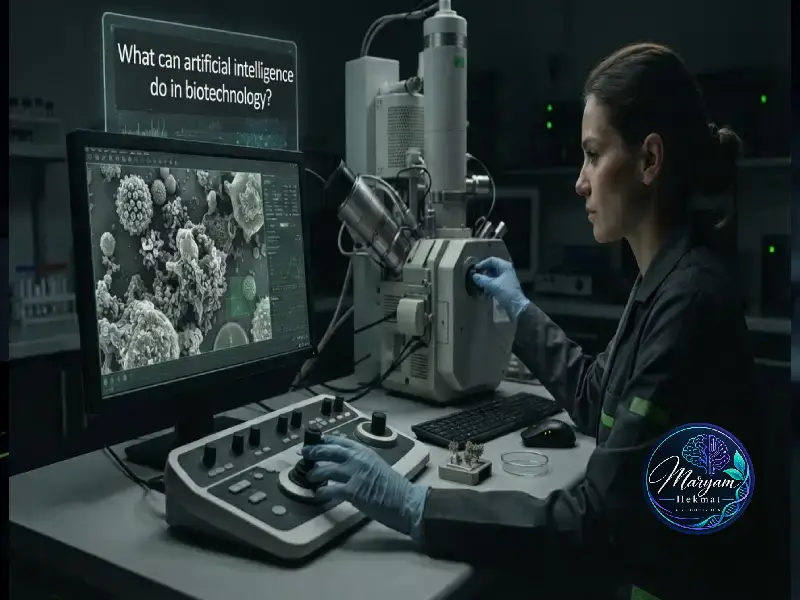
    One of the most important applications of AI in biotechnology is biological image analysis. Microscopy, SEM, and FESEM images contain valuable information about cells, tissues, nanoparticles, and biological structures.

    Computer Vision models can be used for:

    Cell detection and counting

    Image segmentation

    Feature extraction

    Morphological analysis

    Identification of structural changes

    Computer Vision technologies allow researchers to analyze large numbers of images more efficiently.

    Learn more about Computer Vision technologies and their applications in AI-based image processing.


    Example:
    AI-based analysis of SEM/FESEM images can help extract morphological features of nanoparticles and biological structures using image processing and deep learning methods.

    Related Project:
    AI-Based SEM/FESEM Image Analysis

    In this project, deep learning and image processing techniques are used to analyze SEM/FESEM images, extract morphological features, and explore automated analysis of microscopic structures.

    View Project

    How Does Artificial Intelligence Help Biotechnology_


    2. Biological Data Analysis Using Machine Learning

    Biological datasets are often complex and contain multiple variables. Machine Learning algorithms can discover relationships and patterns that may not be easily detected using traditional approaches.

    Applications include:

    Biological sample classification

    Prediction of experimental outcomes

    Genetic data analysis

    Pattern recognition in large datasets

    Access comprehensive bioinformatics databases and computational tools for genomic and biological research:
    National Center for Biotechnology Information (NCBI)


    3. AI in Drug Discovery and Molecular Design

    Drug development is a complex and expensive process. AI can assist researchers by analyzing molecular structures, predicting compound properties, and identifying potential candidates for further investigation.

    However, AI predictions still require experimental validation and laboratory testing.


    Explore the latest research at the intersection of Artificial Intelligence and Biotechnology through Nature Biotechnology, covering advances in machine learning, genomics, bioinformatics, drug discovery, and computational biology.


    Current Applications of AI in Biotechnology

    AI technologies have different levels of maturity. Some applications, such as microscopy image analysis, are already used in research laboratories, while others are still under development.


    Application Area AI Application Current Laboratory Usage
    Microscopy Images Cell detection, counting, and classification Widely used in biological research
    SEM/FESEM Images Particle size, morphology, and structural analysis Research use and automated image analysis development
    Microorganism Detection Identification of bacteria and growth patterns Research and developing applications
    Genetic Data Analysis Pattern discovery and biological prediction Used in bioinformatics research
    Drug Discovery Compound prediction and molecular analysis Growing use in pharmaceutical research
    Protein Design Structure and function prediction Advanced research field
    Smart Laboratories AI combined with robotics and automation Still under development

    The Future of AI in Biotechnology

    Future biotechnology laboratories may combine AI with robotics, sensors, and multiple biological data sources.

    Technology Current Status and Future Direction
    Computer Vision for Biological Images Practical use in research
    3D Biological Image Analysis Developing technology
    Prediction of Cellular Response Still challenging
    AI-Powered Autonomous Laboratories Future development area
    Integration of Genetic and Imaging Data Growing research field

    Conclusion:


    Artificial Intelligence is becoming a powerful tool in biotechnology, helping researchers analyze complex biological data, improve laboratory workflows, and discover new opportunities in life sciences.

    1. Will AI replace biotechnology researchers?

    No. AI is a supportive tool that helps researchers analyze biological data more efficiently and accurately.

    2. Is AI currently used in biological laboratories?

    Yes. AI is used in image analysis, biological data processing, bioinformatics, and drug discovery research.

    3. What is the role of Computer Vision in biotechnology?

    Computer Vision helps analyze biological images, detect cells, classify samples, and extract important visual features.

    4. Do researchers need programming skills for BioAI?

    For developing AI models, skills such as Python, Machine Learning, and data analysis are important.

    5. What are the main challenges of AI in biotechnology?

    Limited high-quality data, model interpretation, and differences between laboratory conditions are major challenges.

    6. Can AI create new drugs?

    AI can support drug discovery by predicting promising compounds, but laboratory experiments are still required.

    7. What career opportunities exist in BioAI?

    BioAI creates opportunities in biological data analysis, medical AI, Computer Vision, and AI-driven research.

  • Real-Time Pill Detection and Color Tracking Using YOLO11 and ByteTrack

    Real-Time Pill Detection and Color Tracking Using YOLO11 and ByteTrack

    Introduction

    Computer vision is becoming increasingly useful in pharmaceutical manufacturing, quality control, and automated inspection systems. Detecting and monitoring pills in real-time can help automate tasks that traditionally require manual inspection.

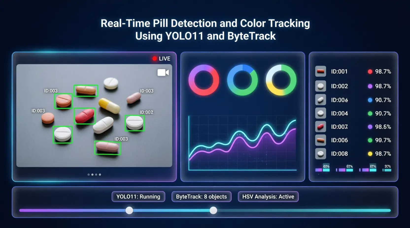
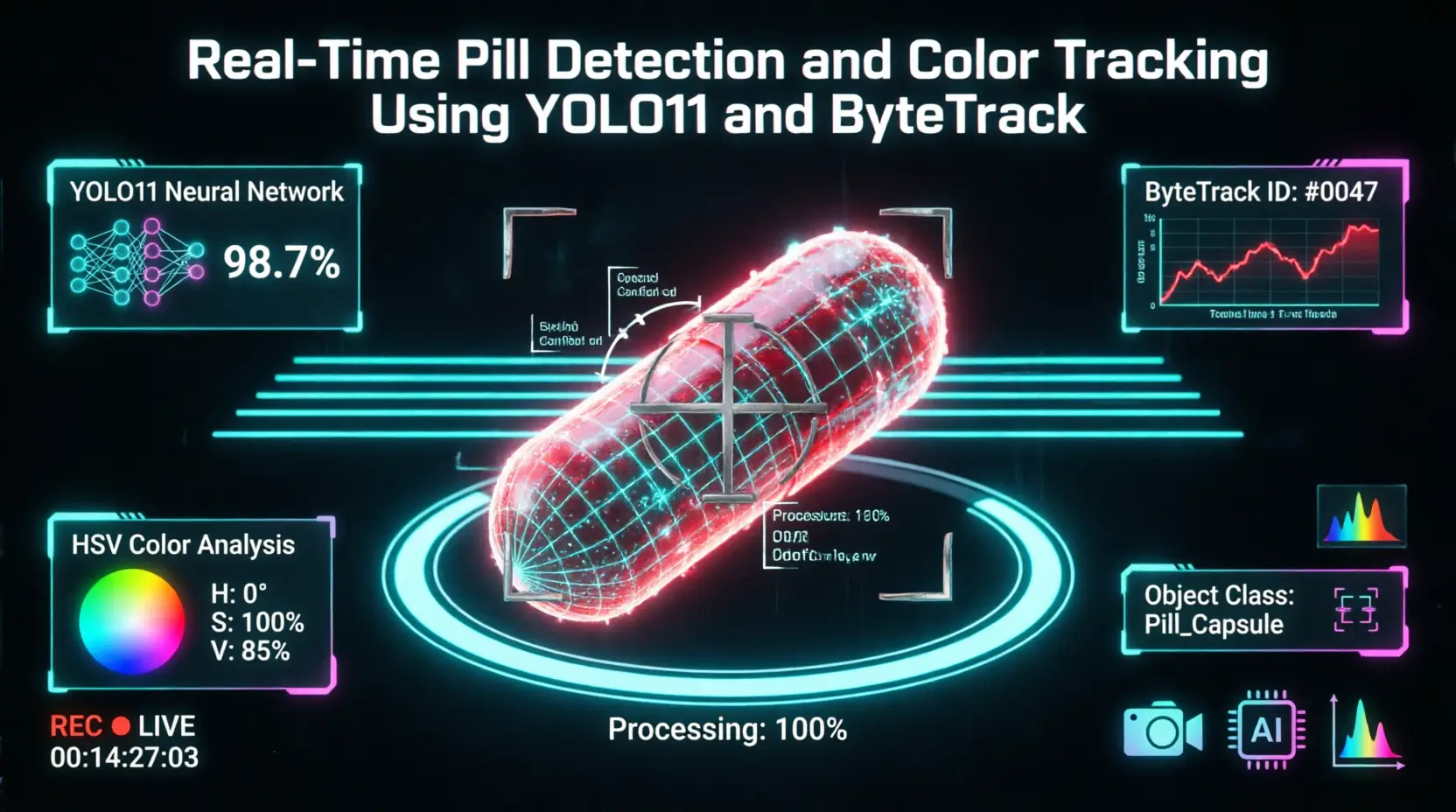
    In this project, I developed a real-time computer vision system for pill detection, object tracking, and color tracking using YOLO11 and ByteTrack.

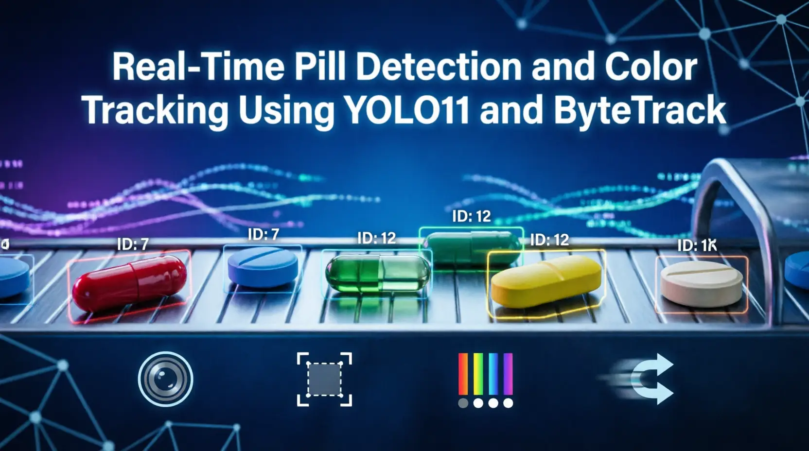
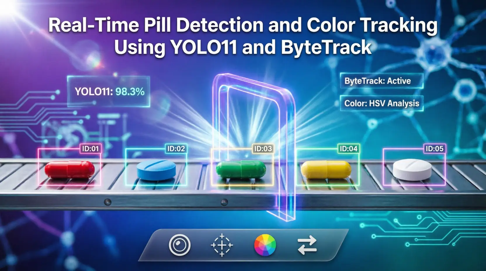
    The system processes video frames, detects pills using a YOLO11 object detection model, assigns persistent IDs to detected objects using ByteTrack, and analyzes their colors during the tracking process.

    Why Pill Detection and Tracking؟

    In pharmaceutical production lines, large numbers of pills may move continuously through a production or inspection area. Manual monitoring can be time-consuming and may be affected by human error.

    An automated vision-based system can continuously monitor the production process and provide useful information such as:

    The number of detected pills

    The position of each pill

    The movement of individual pills

    The color of detected pills

    Potential mismatches or abnormalities

    This makes real-time computer vision a useful approach for developing automated inspection systems.

    YOLO11 for Pill Detection

    The first component of the system is YOLO11, a real-time object detection model developed by Ultralytics.

    YOLO11 can be used for object detection, classification, segmentation, pose estimation, and other computer vision tasks. In this project, the model is used to identify pills in each video frame.

    The official [Ultralytics YOLO11 documentation] provides further information about the model architecture, training, inference, and supported tasks.

    The detection stage provides bounding boxes and confidence scores for the detected pills. These results are then passed to the tracking stage.

    YOLO11 and ByteTrack.

    Object Tracking with ByteTrack

    Detecting an object in every frame is not enough when the goal is to monitor its movement over time.

    For this reason, the project uses ByteTrack for multi-object tracking.

    ByteTrack associates detections across consecutive video frames and assigns a unique tracking ID to each detected object. This allows the system to distinguish between different pills and maintain their identities as they move through the video.

    According to the official [Ultralytics tracking documentation], ByteTrack is a lightweight tracking approach that can be used with YOLO models for multi-object tracking.

    This combination creates a simple but effective pipeline:

    Video → YOLO11 Detection → ByteTrack Tracking → Color Analysis

    Why Pill Detection and Tracking_

    From Biotechnology to Artificial Intelligence: My Journey into AI

    Color Tracking

    In addition to detecting and tracking pills, the system analyzes their colors.

    Color information can be useful when different pills have different visual characteristics. By combining object detection with color analysis, the system can provide additional information about each tracked object.

    The HSV color space is particularly useful for computer vision applications where color needs to be analyzed separately from brightness.

    This approach can make the system more flexible for applications where color classification or color-based monitoring is required.

    Key Features

    The main capabilities of the project include:

    Real-Time Detection

    The system processes video frames continuously and detects pills automatically.

    Multi-Object Tracking

    ByteTrack allows multiple pills to be tracked simultaneously while maintaining individual tracking IDs.

    Color Analysis

    The system can analyze the color characteristics of detected pills and associate this information with tracked objects.

    Automated Monitoring

    Instead of manually monitoring every frame, the computer vision system can continuously analyze the video stream.

    YOLO11 for Pill Detection

    Extensible Architecture

    The system can be extended with additional rules and detection modules for more advanced pharmaceutical inspection applications.

    Potential Applications

    A system like this could be adapted for several industrial applications, including:

    Automated pharmaceutical inspection

    Pill counting

    Color-based pill classification

    Production-line monitoring

    Quality-control assistance

    Detection of mismatched products

    Automated visual inspection

    For example, a future version could combine object detection, tracking, and rule-based analysis to generate an alert when an unexpected pill type, color, or visual condition is detected.

    Benefits of the Approach

    Combining YOLO11 with ByteTrack provides several practical advantages.

    First, object detection and tracking are separated into two clear stages, making the system easier to understand and extend.

    Second, tracking IDs allow the system to analyze the movement of individual pills rather than treating every video frame as an independent image.

    Finally, adding color analysis provides another layer of information beyond simple object detection.

    This combination can serve as a foundation for developing more advanced automated inspection systems.

    Future Improvements

    There are several possible directions for improving the system.

    Future versions could include:

    Pill counting and statistical reporting

    Detection of broken or damaged pills

    Detection of incorrect pill colors

    Automatic alerts for mismatched products

    Improved performance under different lighting conditions

    Integration with industrial cameras and production-line systems

    Deployment on edge devices

    These improvements could turn the current computer vision prototype into a more complete automated inspection solution.

    Conclusion

    This project demonstrates how modern computer vision techniques can be combined to create a real-time pill monitoring system.

    By using YOLO11 for object detection, ByteTrack for multi-object tracking, and color analysis for additional visual information, the system can detect and monitor pills in a video stream while maintaining the identity of individual objects.

    The project also demonstrates how object detection can be combined with tracking and image-processing techniques to build practical computer vision solutions for industrial and pharmaceutical applications.

    Frequently Asked Questions

    1. What is YOLO11 used for in this project؟

    YOLO11 is used to detect pills in each video frame.

    2. Why is ByteTrack used؟

    ByteTrack is used to track detected pills across consecutive frames and maintain their tracking IDs.

    3. Can the system count pills؟

    Yes. The tracking information can be used to implement automated pill counting.

    4. Can the system detect pill colors؟

    Yes. Color analysis can be applied to the detected pills and combined with their tracking information.

    5. Can this project be used in pharmaceutical production؟

    The approach can serve as a foundation for automated visual inspection and can be extended for pharmaceutical production environments.

    Connect With Me

    Interested in my AI and computer vision projects? You can explore more of my work and connect with me through the links below.

    GitHub — Explore my source code and AI projects.

    LinkedIn — Connect with me and follow my professional journey.

    Kaggle — Explore my machine learning experiments and datasets..

  • From Biotechnology to Artificial Intelligence: My Journey into AI

    From Biotechnology to Artificial Intelligence: My Journey into AI

    From Biotechnology to Artificial Intelligence: My Journey into AI

    My experience of changing my career path, learning AI, and entering the world of Computer Vision

    From a Scientific Background to a New Path

    My professional journey began in Biotechnology, a field that taught me how to approach complex problems, work with data, and pay attention to details. Over time, I became increasingly interested in technology and the potential of artificial intelligence to solve problems in different fields.

    As my interest in technology grew, I decided to explore a new path in Artificial Intelligence. What initially started as curiosity gradually became a serious learning journey, leading me to Machine Learning, Deep Learning, and eventually Computer Vision.

    Taking My First Steps in AI

    My first serious step into this field was learning Python. After becoming familiar with programming concepts, I started learning Machine Learning and working with tools and libraries such as NumPy, Pandas, and Scikit-learn. Working on different projects helped me understand how theoretical concepts could be applied to real-world data.

    I then became interested in Deep Learning and Neural Networks. Working with TensorFlow and Keras allowed me to explore different neural network architectures and gain a better understanding of how models learn from data. At this stage, I realized that the best way for me to learn was through practical projects and experimentation.

    پروژه نشخیص ذرات از تصاویر میکرویکپ الکترونی

    Why Computer Vision?

    Among the different areas of Artificial Intelligence, Computer Vision became particularly interesting to me. The ability of a computer system to analyze images and videos, detect objects, and extract useful information from visual data fascinated me.

    This interest encouraged me to learn more about image processing and deep learning models for Computer Vision. From image classification to object detection and video analysis, each project gave me an opportunity to develop my skills and gain more practical experience.

    Working on Real-World Projects

    One of the projects that gave me valuable practical experience was a pill detection, tracking, and color classification system. In this project, I used YOLO-based models to detect pills and tracking techniques to follow them across different video frames. Working on this project allowed me to experience different stages of a Computer Vision system, from model training to video inference and result analysis.

    Projects like this taught me that building an AI system is not simply about choosing a model and training it. Data preparation, model evaluation, debugging, optimization, and finding the right solution for real-world conditions are all important parts of the process.

    Combining Biotechnology and Artificial Intelligence

    My background in Biotechnology remains an important part of my professional journey. I believe that combining knowledge from life sciences with Artificial Intelligence can create interesting opportunities for working on problems related to biology, healthcare, and biological data analysis.

    At the same time, learning AI has allowed me to look at scientific problems from a different perspective. My goal is to eventually combine knowledge from both fields and develop practical solutions where Artificial Intelligence can work alongside biological sciences.

    My Journey Continues

    Learning Artificial Intelligence is not a destination for me; it is an ongoing journey. My current focus is on Machine Learning, Deep Learning, and especially Computer Vision, and I continue to improve my skills by studying new concepts and working on practical projects.

    This article is part of my personal AI journey. Visit my personal website to explore my projects, experience, and other work in Artificial Intelligence and Computer Vision.